Wednesday, November 9, 2011

Microaquarium Madness Act V

Jacob Guillen
11/09/2011
Fourth Post
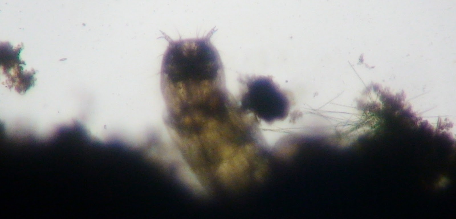
When I observed my Microaquarium under the microscope, there was an even larger decrease of organisms than there was the last observation. The water from the Fountain City Duck Pond was also at a lower level than last time. Despite there being two  Midge Larvae in the aquarium, there were less other organisms in the tank. all of the new organisms that were discovered last week, except the  Oedogonium, ( Nematode, Philodina, and Euchlanis) have all disappeared. Many of the Seed Shrimps and Cyclops are also gone, a sparse few of them are swimming near the sediment at the bottom of the aquarium, scrounging for food. The Vorticella showed no obvious signs of decrease, despite the fact that only two clusters were found. Another discovery, crucial in the explanation of the disappearances of many of the organisms, was that there was no Halteria to be found anywhere. This, along with the fact that many of the other organisms are unable to chew the food, (instead, they just swallow it down), and that the Amblestegium sp. Moss being virtually untouched, leaves me to conclude that the Halteria was the primary food source for many of the organisms in the aquarium. And after the disappearance of the  Halteria, it seemed that a few other organisms could not adapt fast enough to compensate for the disappearance of their food source.

Monday, November 7, 2011

Microaquarium Madness IV

Jacob Guillen
11/03/2011
Blog four
Before putting the micro aquarium under the microscope, I saw that the carnivorous Utricularia gibba L. plant  had all disappeared, except for the stalk at the bottom of the tank, whereas the Amblestegium moss remained unchanged from when it was placed on the left side of the aquarium. When I looked under the microscope, I was dismayed to discover that there were fewer organisms remaining than in the previous entry.   Furthermore, I found the exoskeleton remains of Halteria, Cyclops, and Seed Shrimps littering the entire aquarium. I would also like to clarify that the Vorticella organism was on the Amblestegium moss, rather than the Utricularia gibba L. plant. I sometimes on rare occasion, caught a glimpse of a scurrying Cyclops, or the bounding movement of a Seed Shrimp, however, there was still a dramatic decrease of these organisms overall. The only organism that showed no change in its population, was the Midge Larvae, which was now located around the stem of the Utricularia gibba L. plant. The Halteria and vorticella population also decreased, however, their   decrease was far less drastic than the other organisms. Despite the dropped population of these organisms, new organisms were discovered, and classified, most of which surprisingly appeared after the death of the Utricularia gibba L. plant. The first creature was spotted coming in and out of the decaying carnivorous plant, this was a nematode (unable to determine what genus it is quite yet), which appeared to be motile, as its movement was slowly erratic. Due to its constant interest in the stem of the Utricularia gibba L. plant, it is hypothesized that this nematode type  feeds particularly on dead or dying vegetation, as well as possibly living vegetation. Upon closer inspection, there were were organisms resembling Vorticella except larger. Despite its appearance, it was unrelated, instead it was a part of a different Phylum, called Rotifera, and its genus  is Philodina. It normally is motile, moving in bounds, however, within my aquarium, they seem to be content being sedentary on the  Utricularia gibba L. plant. Its diet consists of plankton and other small organisms that live within its environment. Interestingly enough, there is another organism of the same phylum, called Euchlanis, which is also motile, in both description and in my aquarium, at which it moved at a slow and steady pace while feeding on plankton. Although the  Spirillia algae was nowhere to be found, another algae, Oedogonium, was present. Also capable of photosynthesis, the Oedogonium may be another possible food source for many of the organisms in that environment. The conclusion that I reached, is that many of the decomposers began appearing shortly after the death of the carnivorous Utricularia gibba L. plant, along with smaller organisms, possibly taking advantage of the lack of a large predator, (besides the  Midge Larvae being the predator). My hypothesis for next week's observation,  is that more nematodes may appear, and the Philodina may finally become motile, like the Euchlanis organism.
Oedogonium Algae, Jacob Guillen
Nematode, Jacob Guillen 
Rotifera Euchlanis, Jacob Guillen
Rotifera Philodina, Jacob Guillen
Works Cited


Smith, Gilbert M. The Freshwater Algae of the United States, Second Edition. 1950.
Pennak, Robert W. Freshwater Invertebrates of the United States, Protozoa to Molluska, Third Edition. 1989.


Sunday, October 30, 2011

Micro Aquarium Madness Act III


Jacob Guillen
10/29/11
Blog three
On Friday October 21, 2011, one betta food pellet (produced by Ocean Nutrition, Aqua Pet Americas), which contains fish meal, wheat flower, soy meal, krill meal, minerals, vitamins and preservatives, was placed within the micro aquarium containing water from the Fountain City Duck Pond, attracting many organisms. When I looked into the microscope on 10/25/11, there was an increase in the overall increase in every organism, which leads to my hypothesis that when a food source increases, the overall organism population will increase. I would also like to make a correction, stating that the large microorganisms that were entangled in the carnivorous Utricularia gibba L. were really buds forming on the leaves. However, there seems to be a change in the Utricularia Gibba L. since the last post. The plant seems to be curling into a certain region on the right, while the plant on the left, Amblestegium (moss), seems to be in the same position as it was placed for the first blog. Upon closer observation, I noticed that although there was a general increase in microorganism populations, it was the smaller organisms (x10 magnification) that had  the largest increase. There were an abundance of organisms, called Vorticella attached to, and floating around the carnivorous plant on the right. Vorticella is a single-celled Cilliate, which is mostly sedentary, however, it is able to induce propulsion when necessary. Its natural food source is bacteria (Patterson 1996), which leads me to believe that there are quantities of bacteria around the Utricularia gibba L. plant. Another of the "small" organisms is called Halteria, which is also a single-celled Cilliate, which is found in abundance everywhere within the tank. It moves erratic bounds, free-floating from area to area, and its primary food source is algae (Patterson 1996), although there is only one specimen found at the bottom of the tank. The algae (Spyrogyra), is a sedentary autotroph (Lund ) that is a primary food source for many of the microorganisms in its ecosystem. The larger organisms (x4 magnification) had a relative population growth since last time, although it was not as much as the population of the smaller organisms (x10 magnification). For an indistinct reason, none of these organisms were attracted to light, instead, they made an attempt to keep out of the spotlight as much as possible. One in particular was the Cyclops organism, a free-floating omnivore ( feeds on plant material and dead organisms) which shot from area to area in an erratic motion (Smith 2001) was found intermittent within the tank. Another organism of the same size category is the Seed Shrimp, also a free-floater, it typically moves in the same erratic manner as its fellow organisms. Its primary food sources come from plant material and dead organisms, and on certain occasions, it has been known to feed on smaller organisms as well (Smith 2001), a possible predator to the Vorticella and Halteria organisms. There is one organism, however which is the primary predator to the rest of the medium-sized cells. The Midge Larvae (Chironamidae) is known for its voracious nature in small ecosystems such as these. It is very capable of movement if it wishes (free floater), however, it prefers to lie in wait in the sediment, for unsuspecting prey. As implied, the larvae feeds on other microorganisms, such as the Seed Shrimp and the Cyclops (Ward and Whipple 1918), which would explain the dead exoskeletons scattered around the bottom of the tank. In conclusion, I would  like to hypothesize that there will be another increase in the population of the organisms, except for the midge larvae, depending whether or not the food production increases.


Works Cited
Patterson, D.J. 1996. Free-Living Freshwater Protozoa.
Smith, Douglas Grant.2001. Pennak's Freshwater Invertebrates of the United States, Fourth Edition, Porifera to Crustacea.
Ward, Henry B., Whipple George C. Freshwater Biology. 1918
Lund, Hilda C., John WG. Algae, Their Microscopic World Explored. 
Seed Shrimp, Jacob Guillen


Cyclops, Jacob Guillen

Vorticella, Jacob Guillen
 Midge Larvae, Jacob Guillen
Halteria, Jacob Guillen

Wednesday, October 19, 2011

Microaquarium Madness, Act II

Jacob Guillen
Micro aquarium, second post
10/18/11
Before the experiment began, my hypothesis was that a large amount of small organisms, (ones that can easily be seen by the 10x magnification) would hide around the (carnivorous) Utricularia gibba L. plant on the right side of the tank, in order to obtain protection from larger organisms, (an action similar to that of a clownfish and a sea anemone). since the Utricularia gibba L. plant was carnivorous, I hypothesized that this plant would attack larger organisms, (ones that are able to be seen using the 4x magnification), while the smaller organisms (10x magnification), would dart around the plant, seemingly unaffected by  the carnivorous plant. Meanwhile, in the center and at the left side of the tank, closer to the moss (Amblestegium sp.), there would be less small organisms (10x magnification), and more of the medium and large organisms, (able to be seen using the 4x magnification). However, when I viewed the micro aquarium, I received an interesting surprise, one that exceeded my expectations.

The first change that I noticed immediately, was that my micro aquarium's water supply (from the Fountain City Duck Pond) appeared to be less than last week's amount, possibly hinting that it was being used by the plant materials, and by the microorganisms as well. Around the moss, there were very few signs of microorganisms, while at the center of the tank, there was a small amount of medium-sized organisms (4x magnification), and even less small organisms (10x magnification). However, around the carnivorous Utricularia gibba L. plant, there were many large organisms (at least five organisms), except they all appeared dead, and connected to the carnivorous plant's leaves, while the small organisms seemed plentiful, which raises another hypothesis, one that implies that the smaller organisms (10x magnification) are scavengers feeding on the large dead organisms. Another surprise is that underneath the sediment sample at the bottom of the micro aquarium, there was a microorganism with a likeness very similar to an earthworm, both in shape, and in locomotion. This evokes an assumption that this organism shares the same environmental function as an earthworm, just on a smaller scale. My hypothesis for the following week, is that there will be another decrease in water, and a slight increase in small organisms (10x magnification).

Another curiosity is that when I refilled the micro aquarium with water, the bottle was from an unspecified source, and I am yet unsure whether or not this will affect the experiment in any way.

Thursday, October 13, 2011

Micro Aquarium Madness

Jacob Guillen
Micro Aquarium project Layout:
 The water source that will be used in my experiment is the water from the Fountain City duck pond, (If you wanted to search for the source of the water, you would use the name of the sample, (Fountain City Duck Pond), look it up under google maps, (under Tennessee), and it should be able to give you the adequate location.)  The amount of microorganisms must be determined first by the factors of how they arrived there. First, the water cycle,whether it was transported by natural means, or by human means, the water contained microorganisms, which have been been brought into the new environment. Another factor is by larger animals being used as carriers for the microorganisms, animals such as birds (in general), fish, and any other creatures that used the pond for their basic needs. Next to be explained is the way that the environment supports these little creatures. Larger animals again play a large role in the lives of microorganisms, as they bring and produce food for them, (unintentionally) by decay, defecation, and by carrying small particles along with them. The microorganisms also rely on plants and other microorganisms in order to receive their nourishment.

The experiment requires that a small portion of this water from the pond to be placed in a micro aquarium slide, as well as a soil sample. There are also  two different plants that are to be inserted into the slide. Plant A is Amblestegium, or moss,  hypothetically the food source for many of the microorganisms, will be put in the left side of the slide, whereas, plant B, Utricularia gibba, a flowering carnivorous plant, which may act as a predator to certain microorganisms, will be placed in the right corner of the slide. If you want to know where the plant samples came from, you could do two different techniques of searching. First, you could backtrack to the destinations of the samples before they arrive, (example: Where was Plant A obtained? From a natural spring. Where is it located? Carter Mill Road, and so on.) to make it much easier, you could also use Google maps. Another way to find the plant sample is to look up their habitat range, then make the assumption that it was produced in Tennessee ( if Tennessee is in its range of habitat) because of its convenience of area, (AKA, it is convenient if harvested closer in-state.) then you would  find what habitat it requires to survive, then look up on the map for regions that are capable of creating this environment.  Currently in the slide, there are two microorganisms that I have found, one of which I found while using the 4x magnification, the smaller organism was spotted using the 10x magnification. Now that the experiment is set up, I will review the changes within the following week.